Heights Eye Studio
1533 N Shepherd Dr Ste 120, Houston, TX 77008
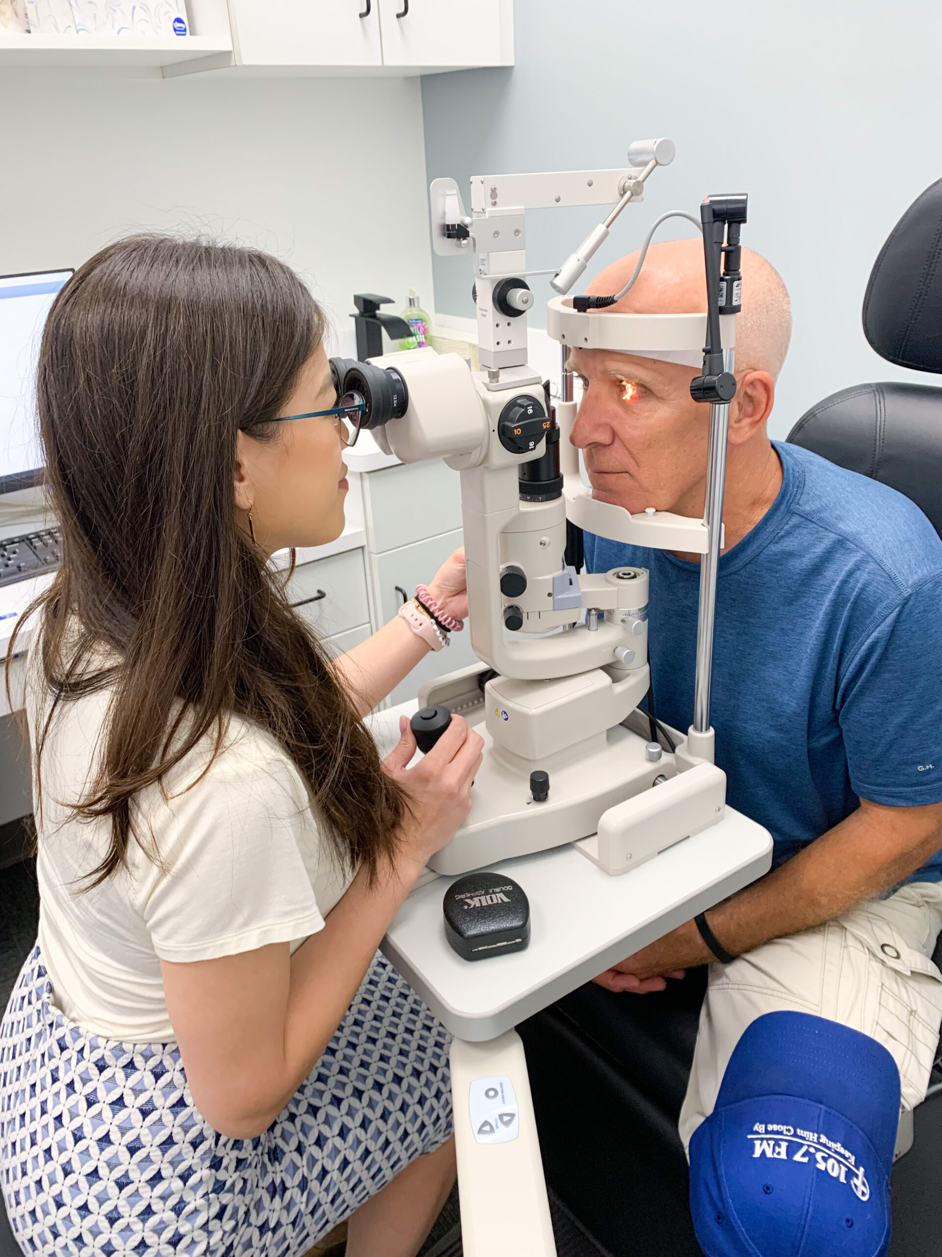
Our Technology
PROKERA
BOOK NOW if interested in trying the Prokera biologic corneal bandages today at Heights Eye Studio
Prokera contains the only FDA-cleared cryopreserved amniotic membrane for anti-inflammatory and anti-scarring. Prokera corneal bandages are designed for treating damaged cornea(s) by creating an environment for regenerative healing. Indications for Prokera include corneal ulcers, keratitis, corneal abrasions, corneal erosions, dry eyes and more.
Healing Vision. Restoring Life.
Early intervention is critical in eye care. Prokera corneal bandages are designed for treating damaged cornea(s) by creating an environment for regenerative healing.
They are easy-to-use and can be inserted in the office by a trained eye care professional. Because Prokera contains cryopreserved amniotic membrane tissue to promote healing, it excels at restoring a normal and healthy epithelium while minimizing the risk of scar tissue formation. Early intervention with Prokera supports the restoration of the cornea’s own healing capabilities, reducing inflammation, improving corneal health, and optimizing long-term outcomes.
Seeing A Difference
Prokera is the only FDA-cleared cryopreserved amniotic membrane product. It supports the corneal healing process without harmful side effects. Our proprietary CryoTek preservation method maintains full biologic components including a key protein complex HC-HA/PTX3, and structural integrity equivalent to fresh tissue, which helps rapidly restore the cornea’s own healing capabilities.
Alternative amniotic membrane solutions that are dehydrated lack crucial biologic components, which risks repeated epithelial defects or erosions, chronic inflammation, scarring, vision loss, or pain.
The Challenge With
Conventional Treatments
Corticosteroids
Increased ocular pressure, cataract formation, and infection risk
Eye Drops
Ocular surface changes, inflammation, and fibrosis. May rely on a single molecule to address inflammation in multi-factorial diseases
Bandage Contact Lenses
No therapeutic benefits, relies on body’s ability to heal itself
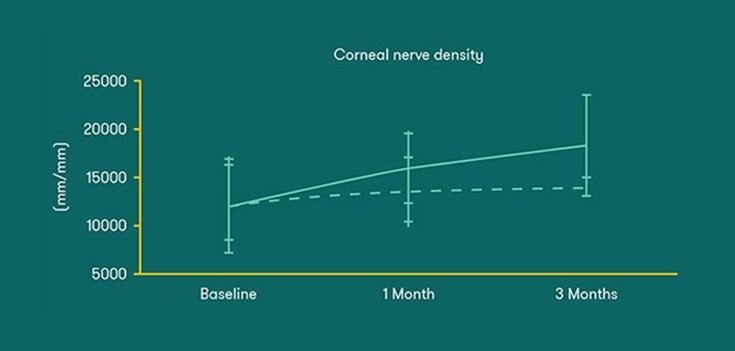
Proven Outcomes
- Improves corneal nerve regeneration and accelerates ocular-surface health recovery
- Shown to significantly improve both corneal nerve density and sensitivity
- Halts fibrosis, facilitating faster re-epithelialization
- Superior outcomes vs bandage contact lens
- — 70% of eyes re-epithelialized by day 5 with Prokera Slim vs. only 20% with bandage contact lens
- — 90% of eyes achieved absolute corneal clarity by Day 7 with Prokera Slim vs. 0% with bandage contact lens
93% Of Patients With Moderate- To-Severe Dry Eye Disease Reported Improvement After One Prokera Treatment.
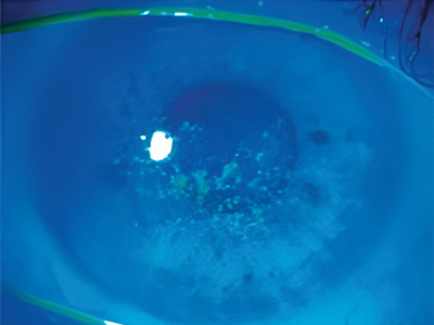
Before Prokera
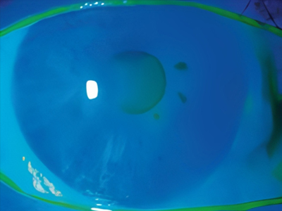
After Prokera
After Prokera photo taken 3 months after treatment. Standard treatment window for Prokera is 3-7 days
The Right Healing.
The Right Product.
We are in a race against time to heal chronic wounds. BioTissue products provide the natural healing properties of human birth tissue to the wound. The BioTissue proprietary CryoTek cryopreservation process is the only tissue processing method shown to produce an allograft comparable to the native tissue.
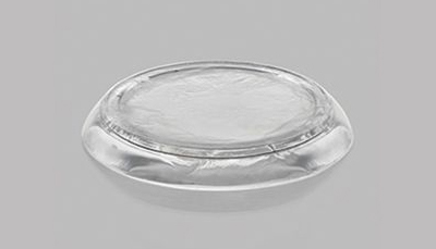
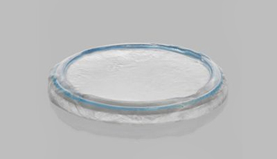
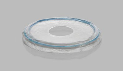
Prokera Slim
Use our ComfortRing™ technology for a lower profile device that contours to the ocular surface to maintain comfort in treatment
More Than 300,000 Prokera Applications
- Corneal ulcer
- Central corneal ulcer
- Corneal ulcer with hypopyon
- Marginal corneal ulcer
- Mycotic corneal ulcer
- Dendritic corneal ulcer
- Neurotrophic keratoconjunctivitis
- Exposure keratoconjunctivitis
- Punctate keratitis
- Filamentary keratitis
- Epithelial corneal dystrophy
- Recurrent corneal erosions
- Band keratopathy
- Nodular corneal degeneration
- Chemical burn
- Thermal burn
- Acid burn
- Steven-Johnson Syndrome
BOOK NOW if interested in trying the Prokera biologic corneal bandages today at Heights Eye Studio!