Heights Eye Studio
1533 N Shepherd Dr Ste 120, Houston, TX 77008
Our Technology
Optomap Daytona
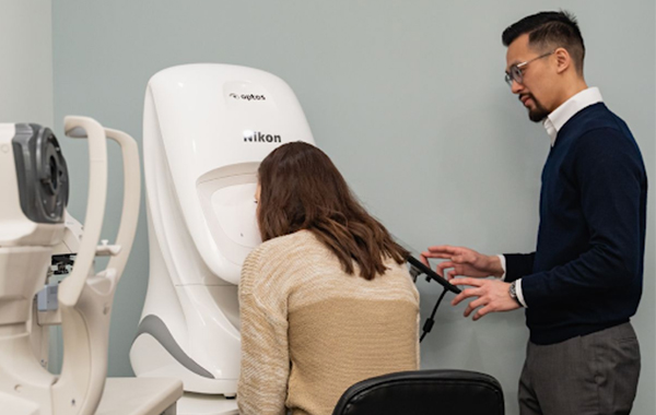
Instead of the dreaded dilation drops that sting and have side effects of blurry vision and light sensitivity, the Heights Eye Studio Experience includes an overall check of the health of your eyes with the new Optomap Daytona. This new piece of state-of-the-art equipment allows us to obtain a 200-degree digital picture of the retina (back of the eye) in order to aid in diagnosing eye diseases. Many asymptomatic eye problems can be found with a quick Optomap photo including retinal holes, retinal tears, retinal detachments, macular degeneration, glaucoma, diabetic retinopathy, and hypertensive (high blood pressure) retinopathy. The Optomap pictures will also be a part of your permanent record on your file, allowing us to compare images a year to year to look for changes.
Daytona produces a 200° single-capture optomap retinal image of unrivaled clarity in less than ½ second. This fast, easy, patient friendly, ultra-widefield imaging technology was designed for healthy eye screening.
Enhances Clinical Decision-making Evaluation of the peripheral retina is critical for optimal patient management.1 optomap imaging is ideal for peripheral examinations. Published studies comparing field of view and clinical utility of various widefield imaging systems confirm optomap captures the widest clinically usable field of view and the most retinal pathology.
Features and Benefits
- Non-mydriatic, non-contact imaging through most cataracts and small (2mm) pupils
- High resolution 200º optomap images improve pathology detection and management from macula through the far periphery
- optomap image clarity yields unrivaled detail across the entire 200° image
- 3-in-1 Color Depth Imaging provides important clinical data from the retinal surface through the choroid
- Autofluorescence imaging (green laser) highlights lipofuscin in the Retinal Pigment Epithelium
- Stereo disc imaging facilitates Optic Nerve Head review
Book here to schedule an eye exam without dilation using the new Optomap Daytona!